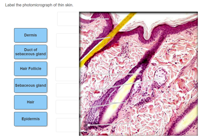
Label the photomicrograph of thin skin – Labeling the photomicrograph of thin skin offers a captivating glimpse into the intricate architecture of our integumentary system. This detailed examination reveals the diverse layers and structures that collectively contribute to the skin’s vital functions.
The epidermis, the outermost layer, serves as a protective barrier against external elements. Beneath lies the dermis, providing structural support and housing essential structures like hair follicles and sebaceous glands. Finally, the hypodermis, the deepest layer, insulates the body and stores energy.
Label the Photomicrograph of Thin Skin

Histological Features of Thin Skin
Thin skin, also known as epidermis, is a type of skin found in areas of the body that are not subjected to excessive friction or pressure. It is characterized by its thinness, flexibility, and high permeability.
The epidermis is the outermost layer of thin skin and consists of multiple layers of keratinized cells. The dermis is the middle layer and contains connective tissue, blood vessels, and nerve endings. The hypodermis is the innermost layer and is composed of adipose tissue.
Labeling the Photomicrograph
The photomicrograph of thin skin shows the following structures:
- Epidermis
- Dermis
- Hypodermis
- Hair follicle
- Sebaceous gland
Significance of Thin Skin
Thin skin is important for:
- Permeability: Thin skin allows for the easy absorption of substances into the body.
- Sensitivity: Thin skin is highly sensitive to touch and temperature.
- Thermoregulation: Thin skin helps the body to regulate its temperature.
Thin skin is found in areas such as the face, neck, and inner arms.
Clinical Applications
Photomicrographs of thin skin are used in dermatology to:
- Diagnose skin conditions
- Monitor treatment progress
- Guide therapeutic interventions
For example, photomicrographs can be used to diagnose skin cancer, monitor the progress of a skin infection, and guide the treatment of a skin condition.
Educational Value, Label the photomicrograph of thin skin
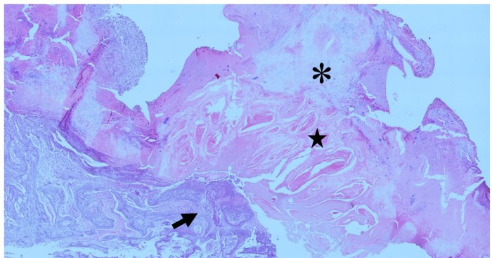
Photomicrographs of thin skin are valuable in teaching histology and anatomy because they provide a clear and detailed view of the skin’s structure.
Photomicrographs can be used to illustrate the different layers of the skin, the cellular composition of each layer, and the relationship between the skin and other structures in the body.
General Inquiries
What are the key anatomical structures visible in a photomicrograph of thin skin?
Epidermis, dermis, hypodermis, hair follicles, sebaceous glands
How does the thickness of thin skin affect its functions?
Thin skin allows for increased permeability, sensitivity, and efficient thermoregulation.
What are some clinical applications of photomicrographs in dermatology?
Diagnosis of skin conditions, monitoring treatment progress, guiding therapeutic interventions


