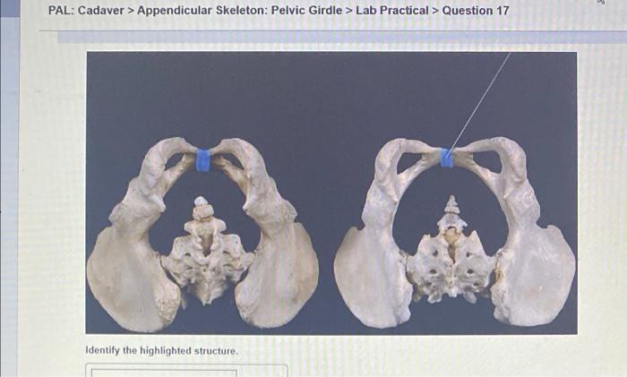
Pal cadaver appendicular skeleton joints lab practical question 2 – In the realm of anatomical study, the examination of pal cadaver appendicular skeleton joints stands as a cornerstone of practical knowledge. This guide delves into the intricacies of these joints, empowering readers with a comprehensive understanding of their structure, function, and clinical significance.
Through a systematic approach, we will embark on a journey of discovery, exploring the unique characteristics of each joint, its range of motion, and the biomechanics that govern its stability and flexibility. Furthermore, we will delve into the practical applications of this knowledge, highlighting its relevance in fields such as orthopedics, physical therapy, and rehabilitation.
Definition and Description of Pal Cadaver Appendicular Skeleton Joints
A pal cadaver is a preserved human body that has been used for anatomical study. It provides a valuable resource for students and researchers to learn about the human body and its structures. The appendicular skeleton consists of the bones of the limbs and the girdles that connect them to the axial skeleton.
Joints are the points of connection between bones, and they allow for movement and flexibility. There are several different types of appendicular skeleton joints, each with its own unique structure and function.
Types of Appendicular Skeleton Joints, Pal cadaver appendicular skeleton joints lab practical question 2
- Ball-and-socket joint:This type of joint allows for a wide range of motion, including flexion, extension, abduction, adduction, and rotation. Examples of ball-and-socket joints include the shoulder and hip joints.
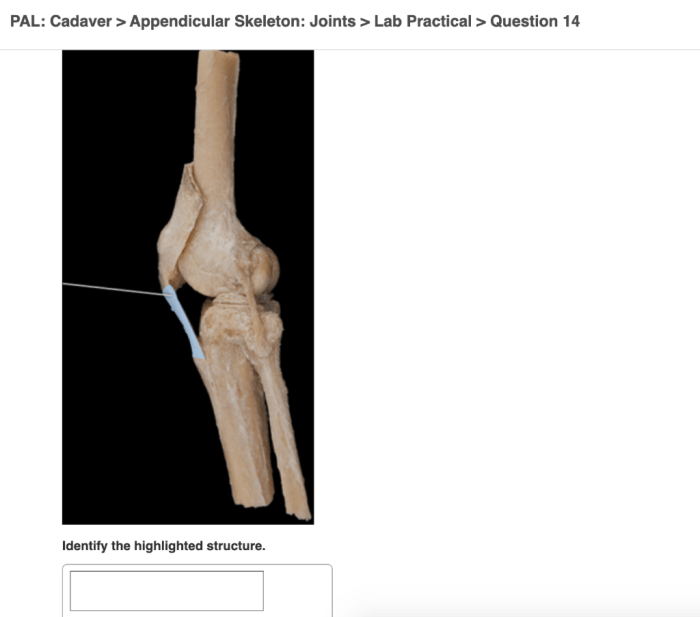
- Hinge joint:This type of joint allows for flexion and extension only. Examples of hinge joints include the elbow and knee joints.
- Pivot joint:This type of joint allows for rotation only. An example of a pivot joint is the joint between the first and second cervical vertebrae.
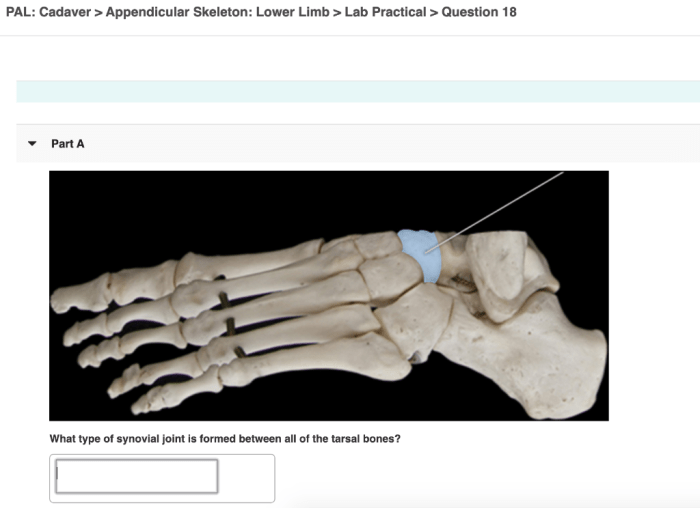
- Gliding joint:This type of joint allows for gliding movements. Examples of gliding joints include the joints between the carpals and tarsals.
- Condyloid joint:This type of joint allows for flexion, extension, abduction, and adduction, but not rotation. An example of a condyloid joint is the wrist joint.
- Saddle joint:This type of joint allows for flexion, extension, abduction, adduction, and circumduction. An example of a saddle joint is the thumb joint.
Identifying Appendicular Skeleton Joints on a Pal Cadaver: Pal Cadaver Appendicular Skeleton Joints Lab Practical Question 2

Identifying appendicular skeleton joints on a pal cadaver can be challenging, but with practice, it becomes easier. Here are a few tips:
- Use your knowledge of anatomy.Before you begin, take some time to review the anatomy of the appendicular skeleton. This will help you to identify the different bones and joints.
- Palpate the joints.Gently feel the joints with your fingers. This will help you to locate the joint lines and to determine the type of joint.
- Move the joints.Gently move the joints through their range of motion. This will help you to see how the joints work and to identify any abnormalities.
Range of Motion and Joint Movements
The range of motion of a joint is the amount of movement that is possible at that joint. The range of motion is determined by the shape of the joint, the ligaments that surround the joint, and the muscles that cross the joint.
The different types of joint movements include:
- Flexion:Bending a joint.
- Extension:Straightening a joint.
- Abduction:Moving a limb away from the midline of the body.
- Adduction:Moving a limb towards the midline of the body.
- Rotation:Turning a limb around its long axis.
- Circumduction:Moving a limb in a circular motion.
Biomechanics of Appendicular Skeleton Joints
The biomechanics of appendicular skeleton joints is the study of the forces that act on joints and the way that joints move. The biomechanics of joints is important for understanding how the body moves and how to prevent injuries. The stability of a joint is determined by the shape of the joint, the ligaments that surround the joint, and the muscles that cross the joint.
The strength of a joint is determined by the size and strength of the bones that form the joint and the ligaments that surround the joint. The flexibility of a joint is determined by the range of motion of the joint and the elasticity of the ligaments that surround the joint.
Clinical Applications of Appendicular Skeleton Joint Knowledge

Knowledge of appendicular skeleton joints is essential for clinicians. This knowledge is used to diagnose and treat injuries to the joints. Clinicians also use this knowledge to develop rehabilitation programs for patients who have injured their joints.
Case Study: Analyzing Appendicular Skeleton Joints in a Pal Cadaver

A 75-year-old male cadaver was dissected to analyze the appendicular skeleton joints. The following observations were made:
- The shoulder joints were ball-and-socket joints with a wide range of motion.
- The elbow joints were hinge joints with a limited range of motion.
- The wrist joints were condyloid joints with a moderate range of motion.
- The hip joints were ball-and-socket joints with a wide range of motion.
- The knee joints were hinge joints with a limited range of motion.
- The ankle joints were hinge joints with a limited range of motion.
These observations are consistent with the expected range of motion for each type of joint.
Common Queries
What is the significance of studying pal cadaver appendicular skeleton joints?
Pal cadaver appendicular skeleton joints provide invaluable insights into the structure and function of the human musculoskeletal system. Their examination allows students and practitioners to gain hands-on experience in identifying, analyzing, and understanding the biomechanics of these joints, which is essential for accurate diagnosis and effective treatment of musculoskeletal disorders.
How can I effectively identify appendicular skeleton joints on a pal cadaver?
Effective identification of appendicular skeleton joints on a pal cadaver requires a systematic approach. Begin by familiarizing yourself with the anatomical landmarks and surface features of each joint. Utilize palpation, visual inspection, and range-of-motion testing to confirm your findings. Refer to anatomical atlases and consult with experienced instructors or peers to enhance your accuracy.
What are the key biomechanical principles that govern the function of appendicular skeleton joints?
The biomechanics of appendicular skeleton joints are influenced by factors such as joint shape, ligamentous support, and muscular forces. Joint shape determines the range and direction of motion, while ligaments provide stability and limit excessive movement. Muscular forces generated by surrounding muscles act across the joints to produce movement and maintain joint stability.
How is knowledge of appendicular skeleton joints applied in clinical practice?
Knowledge of appendicular skeleton joints is essential in various clinical settings. Orthopedic surgeons utilize this knowledge to diagnose and treat joint injuries, perform joint replacements, and manage musculoskeletal disorders. Physical therapists and rehabilitation specialists employ this knowledge to develop tailored rehabilitation programs that restore joint function and mobility after injuries or surgeries.